

RESToRe Lab
Penn Neurosurgery
// Research areas

One question, four diseases, two sets of tools.

The lab works at the boundary between intraoperative neurophysiology, computational analysis, and surgical planning. Hover any acronym below for a quick definition; click any thread heading to jump.

// The through-line

Understanding the neural control of movement, to restore function after neurologic injury.

Functional neurosurgery is one of the few situations where a working human brain can be reached directly. Patients come to the operating room for treatment — a stimulator lead, an electrode to map seizures, a spinal cord stimulator. While they are there, and without changing the operation, we can record how the brain and spinal cord actually produce movement.

The four clinical areas below are separate lines of work. Each has its own reasons, its own collaborators, and its own funding. They are grouped here because they share a setting and a set of tools — not because they test one shared idea. The two method areas are the equipment and software those studies are built on.

// Setting

The operating room

Recordings are added to operations already booked for treatment, and are timed so the operation is not made longer.

// Instruments

High-density arrays and motion capture

Recordings from the surface of the brain, from deep in the brain, and from the spinal cord, matched up with sensors that track how the arms and legs move.

// Software

Capture and analysis tools

Thalamus records everything at once and keeps it in step. nSTAT analyzes what comes out.

// Constraint

Consent and clinical priority

Taking part is always optional, consented separately, and reviewed by the ethics board. It never changes the surgical plan.

// A note on what this page claims

Each section lists what has been published, with a link to the source. Results are given as the papers give them. This page does not draw links between areas that the studies themselves do not make. Where the evidence comes from other groups rather than from this lab, that is said.

Movement disorders — intraoperative neurophysiology during awake deep brain stimulation (DBS).

The lab records single-unit and local-field-potential activity during awake DBS implantation for Parkinson's disease and essential tremor, and uses high-density ECoG arrays in collaboration with Precision Neuroscience.

Work threads:

  • Imaging-guided directional DBS for Parkinson's disease and essential tremor.
  • Cuneiform-nucleus DBS for levodopa-resistant freezing of gait — Phase I pilot, multicenter trial supported by the Michael J. Fox Foundation.
  • MRgFUS for tremor (essential tremor, parkinsonian tremor).
  • Cerebellar DBS for movement disorders in cerebral palsy.
// What we have found so far
  • Three patients aged 14 to 22 with dyskinetic cerebral palsy had stimulating electrodes placed on both sides of the cerebellum, in a part called the dentate nucleus. All three came through the surgery well. Their scores on a standard movement scale improved by 19% to 40%. The authors say the approach looks safe and may help, but that larger studies with longer follow-up are needed before anyone can be sure. Cajigas et al., J Neurosurg 2023 DOI
  • A review paper argues that a small area of the brainstem called the cuneiform nucleus may be a better place to stimulate for freezing of gait than the site tried before, where trials disappointed. This is the reasoning behind the current trial. It is a review of existing work, not a result in patients. Chang, Cajigas et al., Front Syst Neurosci 2020 DOI
Still open: whether cuneiform-nucleus DBS improves levodopa-resistant freezing of gait under blinded, sham-controlled conditions. That is the primary question of the ongoing multicenter phase 1/2 trial (n = 18 across three sites). No results are available yet.

Epilepsy — characterizing the seizure onset zone.

The lab uses SEEG recordings to characterize seizure onset zones in drug-resistant focal epilepsy, including thalamic SEEG and quantitative biomarkers such as neural fragility. Long-term outcomes work tracks LITT for mesial temporal epilepsy.

Work threads:

  • Thalamic SEEG for centromedian and anterior nucleus targets in generalized epilepsy.
  • Quantitative SOZ biomarkers including neural fragility and high-frequency oscillation rates.
  • Long-term outcomes of LITT for mesial temporal epilepsy.
  • Closed-loop responsive neurostimulation (RNS) for multifocal and bitemporal epilepsy.
// What we have found so far
  • Across 268 patients at 11 hospitals, laser treatment of the inner temporal lobe (LITT) left 55.8% free of seizures at one year, and 49.3% by the last check-up — around four years later on average. The authors note this is lower than open surgery achieves, and that open surgery is still a good option for people the laser does not help. Youngerman et al., J Neurol Neurosurg Psychiatry 2023 DOI
  • In 101 patients, looking at how widely abnormal activity was spread between seizures made it easier to tell whether the seizures started in one place or several. Adding it to the existing prediction raised accuracy from 0.68 to 0.79 on a scale where 1.0 is perfect and 0.5 is a coin toss. This was one center only, and the authors call it a step toward a standard tool rather than a finished one. Gallagher et al., Brain Commun 2024 DOI
Still open: whether quantitative markers such as neural fragility localize the seizure onset zone reliably enough to guide a surgical decision. This work is ongoing.

Pain & neuromodulation — refractory back pain and trigeminal neuralgia.

Surgical management of refractory pain and applications of closed-loop neuromodulation. The lab's clinical practice includes SCS, PNS, and VNS applications, plus stereotactic radiosurgery and microvascular decompression for trigeminal neuralgia.

Work threads:

  • Refractory back pain — SCS with closed-loop sensing approaches.
  • Trigeminal neuralgia — microvascular decompression, gamma-knife radiosurgery, and trigeminal nerve field stimulation for refractory cases.
  • Treatment-resistant depression — stereo-EEG workup for closed-loop deep brain stimulation.
// What we have found so far
  • One paper sets out how the team places 10 recording electrodes, five on each side of the brain, as the first step in planning a device for depression that has not responded to other treatment. Brain-imaging maps of nerve fibres are used to aim three of the five. It worked as planned in all three patients reported. This was done at UCSF, before Dr. Cajigas moved to Penn. Starkweather, Sugrue, Cajigas et al., Neurosurgery 2024 DOI
  • A guide to choosing an operation for occipital neuralgia — a nerve pain at the back of the head — when medicines have not helped. It draws on 32 published studies and 11 patients treated here. The authors are clear that the evidence is thin, and that no one operation has been shown to beat the others. That is why the choice is made with the patient rather than for them. Howard et al., Clin Neurol Neurosurg 2023 DOI
Note: the neuromodulation procedures in this track — SCS, DRG, and PNS — are established clinical treatments. The lab's research contribution here is smaller than in the movement and BCI tracks.

Stroke recovery — paired vagus nerve stimulation.

Paired VNS combines an implanted vagus nerve stimulator with intensive rehabilitation for chronic arm and hand weakness after ischemic stroke. During a therapy session the therapist triggers a brief stimulation at the moment of a successful movement attempt.

The lab offers paired VNS at Penn, in a joint evaluation with stroke neurology and rehabilitation medicine. Eligibility, the surgery, and the rehabilitation protocol are described on the patient page.

// Evidence base

The FDA approved paired VNS in 2021, based on a trial called VNS-REHAB (Dawson and colleagues, Lancet 2021). It enrolled 108 people whose stroke was months or years in the past and whose arm was still moderately or severely weak. Neither the patients nor the therapists knew who was getting real stimulation. The people who got it improved about two to three times as much as those who did the same intensive therapy without it. Read the trial.

Brain computer interfaces — volitional control after neurologic injury.

The lab's BCI work spans intraoperative cortical recording with high-density ECoG arrays during essential-tremor focused-ultrasound cases, and a long-running implantable BCI program for hand-grasp restoration in spinal cord injury.

Work threads:

  • Implantable BCI for volitional hand-grasp restoration in cervical SCI (5+-year follow-up published).
  • High-density 1024-channel ECoG decoding of finger / hand kinematics — Precision Neuroscience collaboration.
  • Single-hemisphere bimanual decoding (M.S.E. thesis-level work).
  • Preferential subspace identification (PSID) for movement-speed decoding from M1 ECoG.
// What we have found so far
  • One participant, a 21-year-old man paralysed from the neck down, used a fully implanted brain–computer interface to grip with his hand again. The system read his intention to move and was right about 89% of the time over 29 weeks in the lab, and 88–91% of the time at home. It drove first electrical stimulation of his muscles, then a powered brace. The paper says plainly that it did not test whether this held up over time. Cajigas et al., Brain Commun 2021 DOI
  • A later paper followed the same man for four and a half years. He used the system at home for about 38 minutes a day on average, and it kept working accurately the whole time. The authors say more electrodes and more participants are needed before anyone can say this holds true generally. Davis et al., J Neural Eng 2025 DOI
Still open: the five-year result comes from a single participant. It shows that a chronically implanted BCI can remain stable over years in one person; it does not establish how far that generalizes.
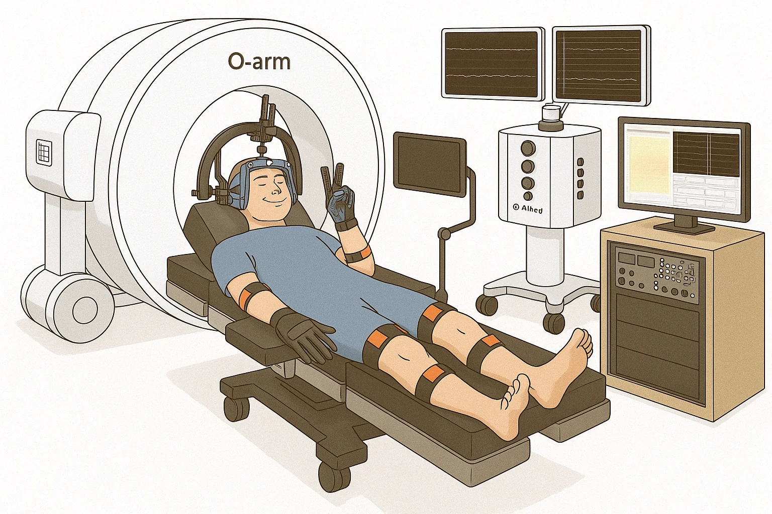
Awake intraoperative recording setup — high-density cortical electrode array with motion-capture gloves
Awake intraoperative setup: 1,024-contact cortical array with motion-capture gloves reconstructing hand and arm movement in 3D. Details on the Clinical Trials page.

Computational methods — tools the rest of the lab runs on.

Open-source software work supporting the experimental program. Thalamus is the lab's closed-loop multimodal data-capture system; nSTAT is the time-series and point-process toolbox.

Work threads:

// Why this thread exists

Thalamus is free, open software that records several kinds of data at once during surgery, and keeps them in step with each other. It takes in brain signals, muscle signals, movement sensors, and pulse oximetry. Tests show it lines these up to better than a thousandth of a second. Haggerty et al., Commun Eng 2026 DOI

nSTAT is a toolbox for analysing the recordings. It fits statistical models to the firing of individual brain cells, tracks how their responses change over time, and works out what a person was doing from their brain activity. It comes in MATLAB and Python. Cajigas, Malik & Brown, J Neurosci Methods 2012 DOI

Both are released openly. See Software for installation, documentation, and citation.
// Funding & collaborators

Supported by

This work is funded in part by the NIH K12 Neurosurgeon Research Career Development Program (NINDS), the Michael J. Fox Foundation for Parkinson's Research (which supports the multicenter trial of cuneiform-nucleus DBS for levodopa-resistant freezing of gait), and the Vazirani Fund for Spinal Cord Research at Penn Medicine, which supports the lab's spinal cord injury BCI program.

Vazirani Fund for Spinal Cord Research · Penn Medicine Penn Neurosurgery